Researchers Expand the Capabilities of Magnetoencephalography

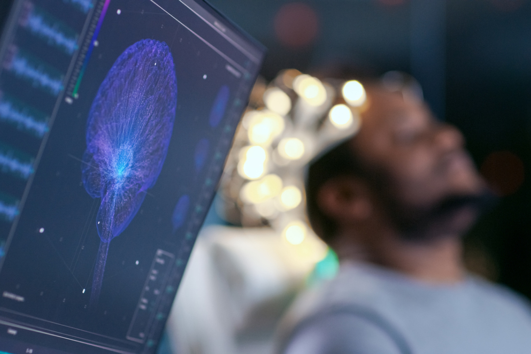
Researchers from the HSE Institute for Cognitive Neuroscience have proposed a new method to process magnetoencephalography (MEG) data, which helps find cortical activation areas with higher precision. The method can be used in both basic research and clinical practice to diagnose a wide range of neurological disorders and to prepare patients for brain surgery. The paper describing the algorithm was published in the journal NeuroImage.
Magnetoencephalography (MEG) is a method based on measuring very weak magnetic fields (several orders of magnitude weaker than the Earth’s magnetic field) induced by the brain's electrical activity. When using MEG, researchers face the complicated task of understanding which areas inside the brain were active when they only have the measurements of sensors placed around the head. This problem is called an ‘inverse problem’ and fundamentally has no universal solution: any set of measurements can be explained by an endless number of different configurations of neural activity sources on the cortex.
To make application of MEG practical, special mathematical methods are used to turn sensor signals into cortical activity maps. These methods can be categorized into two groups. As part of the so-called ‘global’ approach, the multitude of possible solutions for the inverse problem is narrowed down based on the generalized a priori assumptions on brain activity. Under these constraints researchers look for a distribution of sources in the cortex that would explain the measured data. The ‘local’ methods, including the algorithm described in the paper (ReciPSIICOS), aim to find separate sources, and only after that, to create a complete image of brain activity.
ReciPSIICOS uses adaptive beamformers (BF) – a method to process sensor measurements that allows detection of an activity signal of a target neuronal population. For this purpose, it attempts to mute the signals from other sources, but not from all of them as is done in the ‘global’ approach, but instead only the ones that are active at the moment.
When suppressing only active signals, this approach is able to provide a considerably higher fidelity in activity visualization as compared to the ‘global’ approach. However, this method can also suppress the target signals begotten by neuronal ensembles activated simultaneously with neuronal populations in other brain areas. In real-life conditions, such correlation reflects the interaction between neuronal populations, which is an inherent property of the brain, and researchers have to look for methods to overcome this obstacle.
Information on active neuronal populations and the nature of their interaction is encoded in a special covariance matrix, which can be calculated based on the sensor data. This matrix is used by the beamforming algorithm to decide which of the sources should be suppressed. Strictly speaking, this approach is applicable only when sources do not interact: information on such interaction is also contained in the correlation matrix and negatively impacts beamforming algorithm performance. Using the observed data model and the correlation matrix model, the researchers developed a mathematical algorithm that is able to erase the information on sources’ interaction from the correlation matrix. This way they extended the range of applicability of the beamforming method to the environment with synchronous neuronal sources and provided the necessary precision in the visualization of interacting neuronal populations.

Alexey Ossadtchi,
Ph.D., Director of the HSE Centre for Bioelectric Interfaces, the author of the new methods
Magnetoencephalography technology combines the ability to register precise aspects of the temporal evolution in neuronal activity and a potentially high fidelity of localizing the active neuronal populations. The first feature comes from registration of electrical activity that is changing significantly faster than the hemodynamic responses exploited by fMRI, a popular functional brain imaging modality. To achieve a high precision in spatial localization complicated mathematical methods are needed. The family of ReciPSIICOS and PSIICOS methods is an example of mathematical algorithms aimed at increasing the spatial resolution of MEG modality detect active and interacting neuronal populations.
To evaluate the algorithm performance, the researchers first generated a dataset that mimics the signals received by the sensors in real-life and tested four methods on it: two types of ReciPSIICOS and two previously developed algorithms (linearly constrained minimum variance (LCMV) beamformers, and Minimum-Norm Estimates (MNE) approach). In situations when there is no correlation between signals, LCMV and both ReciPSIICOS methods work well, but when there is a correlation, ReciPSIICOS handles the task much better than its predecessors. Under the stress test for the forward modelling accuracy the results are similar: ReciPSIICOS proved to be less sensitive to inaccuracy of the models used, which are inevitable in practice. The scholars also demonstrated operability and high performance characteristics of the new approach on several real MEG datasets characterized by the presence of synchronous neuronal sources that could not be adequately processed by the classical beamforming algorithm.
See also:
'Neurotechnologies Are Already Helping Individuals with Language Disorders'
On November 4-6, as part of Inventing the Future International Symposium hosted by the National Centre RUSSIA, the HSE Centre for Language and Brain facilitated a discussion titled 'Evolution of the Brain: How Does the World Change Us?' Researchers from the country's leading universities, along with health professionals and neuroscience popularisers, discussed specific aspects of human brain function.
‘Scientists Work to Make This World a Better Place’
Federico Gallo is a Research Fellow at the Centre for Cognition and Decision Making of the HSE Institute for Cognitive Research. In 2023, he won the Award for Special Achievements in Career and Public Life Among Foreign Alumni of HSE University. In this interview, Federico discusses how he entered science and why he chose to stay, and shares a secret to effective protection against cognitive decline in old age.
'Science Is Akin to Creativity, as It Requires Constantly Generating Ideas'
Olga Buivolova investigates post-stroke language impairments and aims to ensure that scientific breakthroughs reach those who need them. In this interview with the HSE Young Scientists project, she spoke about the unique Russian Aphasia Test and helping people with aphasia, and about her place of power in Skhodnensky district.
Neuroscientists from HSE University Learn to Predict Human Behaviour by Their Facial Expressions
Researchers at the Institute for Cognitive Neuroscience at HSE University are using automatic emotion recognition technologies to study charitable behaviour. In an experiment, scientists presented 45 participants with photographs of dogs in need and invited them to make donations to support these animals. Emotional reactions to the images were determined through facial activity using the FaceReader program. It turned out that the stronger the participants felt sadness and anger, the more money they were willing to donate to charity funds, regardless of their personal financial well-being. The study was published in the journal Heliyon.
Spelling Sensitivity in Russian Speakers Develops by Early Adolescence
Scientists at the RAS Institute of Higher Nervous Activity and Neurophysiology and HSE University have uncovered how the foundations of literacy develop in the brain. To achieve this, they compared error recognition processes across three age groups: children aged 8 to 10, early adolescents aged 11 to 14, and adults. The experiment revealed that a child's sensitivity to spelling errors first emerges in primary school and continues to develop well into the teenage years, at least until age 14. Before that age, children are less adept at recognising misspelled words compared to older teenagers and adults. The study findings have beenpublished in Scientific Reports .
Meditation Can Cause Increased Tension in the Body
Researchers at the HSE Centre for Bioelectric Interfaces have studied how physiological parameters change in individuals who start practicing meditation. It turns out that when novices learn meditation, they do not experience relaxation but tend towards increased physical tension instead. This may be the reason why many beginners give up on practicing meditation. The study findings have been published in Scientific Reports.
Processing Temporal Information Requires Brain Activation
HSE scientists used magnetoencephalography and magnetic resonance imaging to study how people store and process temporal and spatial information in their working memory. The experiment has demonstrated that dealing with temporal information is more challenging for the brain than handling spatial information. The brain expends more resources when processing temporal data and needs to employ additional coding using 'spatial' cues. The paper has been published in the Journal of Cognitive Neuroscience.
Neuroscientists Inflict 'Damage' on Computational Model of Human Brain
An international team of researchers, including neuroscientists at HSE University, has developed a computational model for simulating semantic dementia, a severe neurodegenerative condition that progressively deprives patients of their ability to comprehend the meaning of words. The neural network model represents processes occurring in the brain regions critical for language function. The results indicate that initially, the patient's brain forgets the meanings of object-related words, followed by action-related words. Additionally, the degradation of white matter tends to produce more severe language impairments than the decay of grey matter. The study findings have been published in Scientific Reports.
New Method Enables Dyslexia Detection within Minutes
HSE scientists have developed a novel method for detecting dyslexia in primary school students. It relies on a combination of machine learning algorithms, technology for recording eye movements during reading, and demographic data. The new method enables more accurate and faster detection of reading disorders, even at early stages, compared to traditional diagnostic assessments. The results have been published in PLOS ONE.
HSE University and Adyghe State University Launch Digital Ethnolook International Contest
The HSE Centre for Language and Brain and the Laboratory of Experimental Linguistics at Adyghe State University (ASU) have launched the first Digital Ethnolook International Contest in the Brain Art / ScienceArt / EtnoArt format. Submissions are accepted until May 25, 2024.


